Our Office
Visit Us Online

At Frankford Dental Group, we use advanced imaging tools to enhance diagnostic confidence and patient outcomes. One of the cornerstones of our diagnostic suite is cone-beam computed tomography (CBCT), which provides three-dimensional views our dentists rely on to plan treatments accurately and safely. CBCT complements clinical exams and conventional radiographs by revealing anatomic detail critical to successful results.
This page explains how CBCT works, why it matters in modern dental care, and the ways we integrate three-dimensional imaging into patient care. Our goal is to provide clear, practical information so you can understand how CBCT supports precise diagnosis and treatment planning.
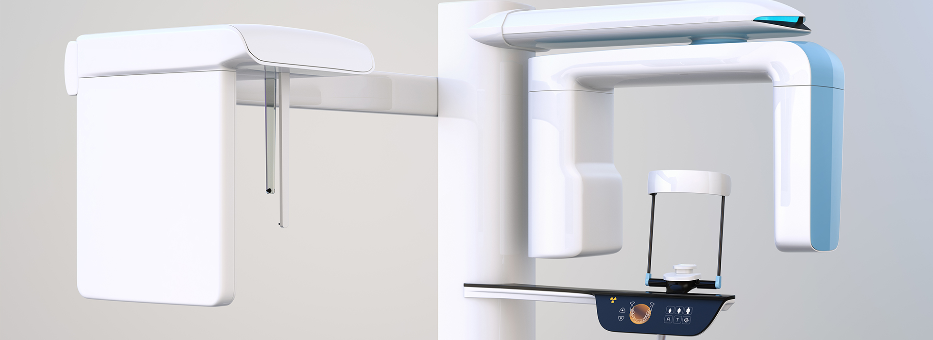
Cone-beam computed tomography uses a cone-shaped X-ray beam and a flat-panel detector to capture a series of images while the scanner rotates around the head. These images are reconstructed into a 3D volumetric dataset that can be viewed in multiple planes. The result is a compact, high-resolution model of teeth, jaws, sinuses, nerve canals, and adjacent structures.
Unlike traditional two-dimensional X-rays, CBCT removes overlaps and distortions that can obscure important details. Clinicians can examine cross-sections or generate virtual slices at any angle, making it easier to locate hidden pathology, evaluate bone volume, and assess spatial relationships before treatment. This flexibility is especially helpful when standard radiographs are inconclusive.
Modern CBCT systems are designed specifically for dental use, with field-of-view options that limit exposure to the area of interest. This targeted approach reduces radiation relative to larger medical CT scans while delivering the detail dentists need for precise planning. The technology balances diagnostic value and patient safety, enabling better-informed decisions.
CBCT is invaluable for implant planning. Successful implant placement requires understanding bone shape, density, and the location of vital structures such as the inferior alveolar nerve and maxillary sinus. CBCT provides a three-dimensional perspective that allows precise implant mapping.
By visualizing the jaw in 3D, our team can simulate implant placement virtually, select optimal implant sizes, and determine if bone grafting or sinus modification is necessary. This preoperative insight reduces surprises during surgery and supports restorative-driven planning, ensuring implants are positioned where the final crown or bridge will function and look best.
CBCT also aids complex extractions, surgical exposure of impacted teeth, and orthognathic assessments. The detailed anatomy captured informs the sequence of care, helps anticipate complications, and supports a more efficient workflow from diagnosis through follow-up.
CBCT extends beyond implant dentistry. Endodontists use 3D imaging to detect root fractures, accessory canals, and periapical pathology that may not appear on 2D films. Oral surgeons rely on CBCT to evaluate cysts, pathology, and lesion relationships.
Orthodontic and airway assessments also benefit. CBCT reveals airway volume and narrowings that may contribute to sleep-disordered breathing evaluations. Orthodontists use 3D data to assess tooth position, jaw asymmetries, and growth patterns when planning comprehensive movement or surgical-orthodontic cases.
Periodontists and restorative dentists use CBCT to measure bone levels, evaluate furcation involvement, and plan ridge preservation or augmentation. In every specialty, a complete three-dimensional map improves diagnostic accuracy and makes treatment recommendations more predictable.
Patient safety is a top priority. Dental CBCT units minimize exposure by limiting the scanned volume and using optimized protocols. The expected diagnostic benefit is always weighed against the radiation dose.
Our practice follows contemporary imaging guidelines and dose-reduction strategies. We choose the smallest field of view that answers the clinical question and adjust settings for each patient’s size and evaluation area. When conventional radiographs provide sufficient information, we use those instead of CBCT.
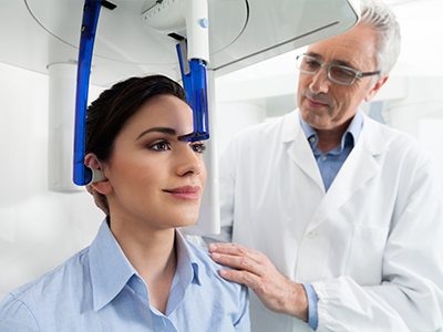
From a patient perspective, CBCT scans are quick, noninvasive, and typically completed in under a minute. The open design is more comfortable than enclosed medical CT scanners, with no injections or intravenous contrast required for routine imaging.
Generating a 3D scan is only the first step; accurate interpretation is essential. Our clinicians review CBCT datasets with specialized software for measurements, simulated implant placement, and anatomy assessment from multiple angles. This transforms raw data into actionable information.
When findings suggest issues outside the typical dental scope—such as suspicious soft-tissue lesions or conditions requiring medical evaluation—we coordinate care with specialists. CBCT helps us recognize when collaboration or referral is in the patient’s best interest, ensuring comprehensive management.
We also use CBCT results to enhance patient communication. Visualizing anatomy in 3D helps patients understand recommended procedures, potential risks, and expected outcomes, supporting informed decision-making and a collaborative approach to care.
Incorporating CBCT into our diagnostic toolkit allows Frankford Dental Group to deliver treatment plans that are precise, predictable, and tailored to each patient's anatomy. If you have questions about whether CBCT is appropriate for your situation or how it would be used in your care, please contact us for more information. We are happy to discuss how three-dimensional imaging may help support your treatment goals.
CBCT, or cone beam computed tomography, is an advanced imaging method that captures three-dimensional images of the teeth, jaws and surrounding structures. The scanner rotates around the head to create a volumetric data set that clinicians can view in multiple planes and cross-sections. This 3-D perspective reveals spatial relationships and anatomical details that are not visible on standard two-dimensional radiographs.
Traditional dental x-rays produce flat, two-dimensional images that are useful for routine exams and detecting cavities, but they can obscure overlapping structures and depth information. CBCT provides precise measurements of bone height, width and density as well as the position of nerves and sinuses, which improves diagnostic accuracy for complex cases. For many advanced procedures, that additional information leads to safer, more predictable treatment planning.
A dentist may recommend a CBCT scan when detailed 3-D information is needed to diagnose a problem or plan treatment that cannot be adequately assessed with standard x-rays. Common reasons include planning dental implant placement, evaluating complex tooth anatomy, assessing impacted teeth, mapping jaw pathology, and guiding oral surgery. The scan helps clinicians visualize critical structures like nerve canals and the sinus floor to minimize intraoperative risks.
At Frankford Dental Group we use CBCT selectively to support clinical decision-making and to improve outcomes for procedures that benefit from volumetric imaging. Your dentist will weigh the diagnostic benefits against the limited additional radiation exposure and recommend CBCT only when it meaningfully changes the treatment approach. If a scan is advised, the team will explain how the information will be used in your specific case.
CBCT exposes patients to a level of radiation that is generally higher than a single traditional dental x-ray but lower than most medical CT scans, and modern protocols minimize dose while preserving image quality. Clinicians adhere to the ALARA principle—keeping radiation "as low as reasonably achievable"—by selecting the smallest field of view and lowest exposure settings appropriate for the diagnostic task. Protective measures, such as thyroid collars and limiting the scanned region, further reduce unnecessary exposure.
Before ordering a CBCT scan, your dentist will consider your medical history, age and the clinical necessity of the study to ensure benefits outweigh risks. Pregnant patients should always inform the dental team, as routine dental imaging is typically deferred during pregnancy unless absolutely necessary. If you have concerns about radiation, the staff can review dose comparisons and explain why CBCT is the most appropriate option for your situation.
Preparation for a CBCT scan is minimal and usually does not require any special steps aside from arriving with removable metal objects taken out of the head and neck area. You will be asked to remove eyeglasses, jewelry, hairpins and any removable dental appliances that could create artifacts in the image. Wearing comfortable clothing and following the receptionist's arrival instructions will help keep the appointment efficient.
Because the scan is noninvasive and quick, there is typically no need for fasting, medication changes or sedation unless your dentist has indicated otherwise for a specific clinical reason. If you have mobility limitations or anxiety, let the team know in advance so they can make accommodations to ensure your comfort during image acquisition. The technologist will position you and confirm you remain still for a short rotational scan that usually takes less than a minute.
CBCT provides comprehensive 3-D views that allow clinicians to assess bone architecture, root morphology, canal configurations, impacted tooth positions and the spatial relationship of anatomical landmarks with high precision. This capability is especially valuable for identifying accessory canals, fractures, root resorption and the proximity of planned implants to nerves or sinuses. It also aids in detecting pathologic lesions and evaluating jaw joint disorders with greater detail than planar radiographs.
Because CBCT captures volumetric information, it enables virtual measurements and cross-sectional slices that guide surgical planning and fabricating surgical guides. These insights reduce surprises during treatment and enable more accurate, individualized approaches for complex restorative, endodontic and orthodontic cases. In short, CBCT translates hidden anatomic complexity into actionable diagnostic information.
A typical CBCT appointment is brief; the actual scan acquisition commonly takes less than one minute, though total appointment time may be 10 to 20 minutes to allow for positioning and review. During the scan you will stand or sit while the machine rotates around your head, and the operator will ask you to remain still and to hold your bite or head in a specified position. The procedure is painless and does not require injections or contrast agents for routine dental applications.
After the images are captured, the dentist or radiologist will reconstruct the volume and review multiplanar views to interpret the scan. Depending on the complexity, the clinician may discuss preliminary findings that day and explain how the CBCT images will influence your treatment plan. If additional specialist input is needed, the images can be shared electronically for collaborative review.
CBCT is instrumental in implant planning because it provides exact measurements of bone height, width and density, and it clearly shows the location of critical structures such as the inferior alveolar nerve and maxillary sinus. By analyzing cross-sectional slices and three-dimensional models, clinicians can select the optimal implant position, angulation and length to achieve stable support while avoiding anatomic hazards. This information supports safer surgery and improves the predictability of restorative outcomes.
Many practices use CBCT data to design surgical guides or to perform virtual implant placement that is later translated into the clinic. The combination of CBCT imaging and digital planning minimizes guesswork and can reduce surgery time and postoperative complications. Your dentist will explain how CBCT findings influence implant selection and the staged sequence of care for your case.
Yes, CBCT is a valuable tool in endodontics for evaluating complex root canal systems, detecting missed canals, identifying vertical root fractures and assessing periapical pathology that may be obscured on conventional x-rays. The ability to view teeth in three dimensions helps clinicians locate additional canals and understand canal curvature and bifurcations, which is critical for successful root canal therapy. CBCT can also reveal the extent of infection and its relationship to surrounding bone and adjacent teeth.
CBCT is typically reserved for cases where traditional imaging is inconclusive or when retreatment, surgical endodontics or complex anatomy is suspected. The dentist will determine whether the added diagnostic detail will change the clinical approach. When used appropriately, CBCT can improve the accuracy of diagnosis and the effectiveness of endodontic treatment.
CBCT should be used judiciously and is not indicated for every patient or routine screening. Pregnant individuals are generally advised to avoid elective dental imaging, including CBCT, unless an urgent clinical need exists and the benefits clearly outweigh risks. Additionally, clinicians consider patient age, prior imaging, and the clinical question at hand; for many routine assessments, conventional radiography remains sufficient and preferable.
Patients with difficulty remaining still or severe claustrophobia may require special accommodations to obtain useful images, and the dental team will assess alternatives as needed. If you have concerns about whether CBCT is appropriate for you, discuss your medical history and specific diagnostic needs with your dentist so the safest and most informative imaging strategy can be chosen.
CBCT scans are performed in-office at many dental practices that have invested in the equipment and training, and they may also be obtained at imaging centers that specialize in dental radiology. In Lubbock, scans can be completed at the practice location to streamline planning and consultations, and scans captured off-site are typically transmitted electronically for review. The technologist or dental assistant will position you and operate the scanner under the dentist's supervision.
The resulting images are interpreted by the treating dentist and, when appropriate, by a board-certified oral and maxillofacial radiologist or other specialists who provide a formal read. Collaborative interpretation ensures that subtle findings are recognized and that the imaging informs a safe, effective treatment plan. If you would like a copy of your images or a specialist consultation, ask the office staff about obtaining and sharing the CBCT data for further review.
Available after hours by appointment.
